
Figure 1.
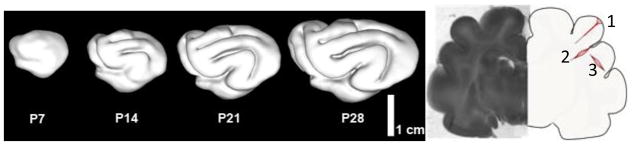
Summary of cortical folding studies in the ferret (4, 5, 21, 49). (a) Sequence of cortical surfaces generated from longitudinal MR imaging studies in the neonatal ferret. PXX = XX days after birth. (b) Coronal slice of P18 ferret brain near the conclusion of the folding process. The illustration on the right summarizes the results of dissection studies of tissue stress (21). Initial cuts (dotted lines) open when tension is normal to the cut. 1: radial cuts through gyri stay closed (showing lack of tension between gyral walls) except at outer surface, where circumferential tension exists. 2: radial cuts through the bases (fundi) of sulci open in subcortical layers, showing circumferential tension in these locations. 3: circumferential cuts through gyri open, showing radial tension along the axes of gyri.