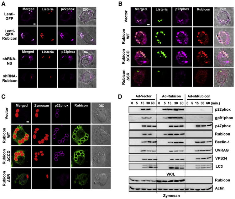
Figure 3. Rubicon Affects p22phoxPhagosome Recruitment and Phagocytosis.
(A–C) (A) Expression or depletion of Rubicon affects the colocalization of p22phox with L. monocytogenes-containing phagosomes. At 48 hr postinfection with lenti-GFP or lenti-GFP-Rubicon (top) at MOI =100 (Figure S3A) or with lenti-shRNA-NS or lenti-shRNA-Rubicon (bottom) at MOI =50 (Figure S3B), Raw264.7 cells were infected with HK-TRITC-labeled L. monocytogenes (MOI = 1) for 30 min, followed by confocal microscopy with αp22phox. Bar, 2 μm. Rubicon enhances the colocalization of p22phox with L. monocytogenes (B) or zymosan particles-containing phagosomes (C) in a p22phox-binding-dependent manner. Raw264.7 cells containing vector, Rubicon WT, ΔCCD or ΔSR were infected with HK-GFP-L. monocytogenes (B) or Texas red-labeled opsonized-zymosan particles (C) for 30 min, followed confocal microscopy with αp22phox or αFlag. Bar, 2 μm.
(D) Rubicon enhances phagocytosis in a p22phox-binding-dependent manner. At 48 hr postinfection with recombinant Ad-Vector (MOI = 200), Ad-Rubicon (MOI = 100), or Ad-shRubicon (MOI = 200) virus, Raw264.7 cells were stimulated with zymosan-coated particles for indicated times, followed by lysis and sucrose-gradient ultracentrifugation to isolate the bead-containing phagosomal fractions. Phagosomal fractions were subjected to IB with αBeclin-1, αUVRAG, αgp91phox, αp22phox, αp47phox, αVPS34, αLC3 or αRubicon. WCL were used for IB with αRubicon or αActin. See also Figure S3.

