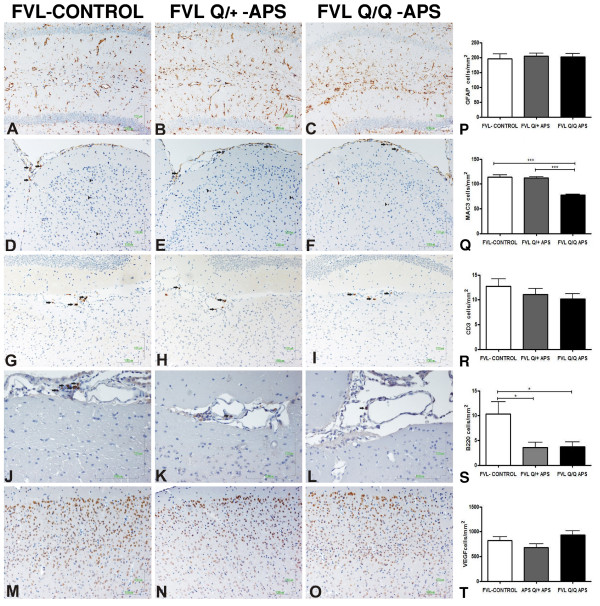
Figure 4.
Immunohistochemical staining for inflammatory and vascular markers in factor V Leiden (FVL) mice. Representative immunohistochemical staining images from the three groups: adjuvant-immunized FVL control (FVL-control), experimental antiphospholipid syndrome (eAPS), heterozygous FVL (FVLQ/+-APS) and eAPS homozygous FVL (FVLQ/Q-APS) mice. Quantification data for each marker are also presented. (A–C,P) Glial fibrillary acidic protein (GFAP)-positive immunoreactions with similar expression in the area of the hippocampus (original magnification × 20). (D–F,Q) MAC3-positive cells (macrophages) in the meninges (black arrows) and in the parenchyma of the cortex (black arrowheads; original magnification × 20). (G–I,R) CD3-positive cells (T cells, black arrows; original magnification × 20). (J–L,S) Infiltrates with increased expression of B220-positive cells (B cells) in the control FVL group compared with the APS FVLQ/+ and APS FVLQ/Q groups (black arrows; original magnification × 40). (M–O,T) Representative images of vascular endothelial growth factor (VEGF) staining, with similar expression in the area of the cortex (original magnification × 20).

