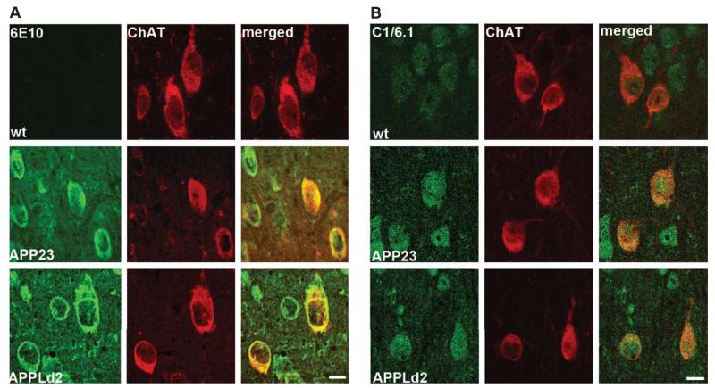
Fig. 2.
Basal forebrain cholinergic neurons from APP23 and APPLd2 mice overexpress human AβPP. A) Co-immunolabeling for ChAT (red) and antibody 6E10 (green), which reacts with human AβPP, βCTFs, and Aβ, in the MSN of APP23 (middle panels) and APPLd2 (bottom panels) mouse brain tissue. No 6E10 immunolabeling is seen in the wt mice (top panels). B) Co-immunolabeling for ChAT (red) and antibody C1/6.1 (green) [25], which reacts with human and murine AβPP/αCTF/βCTF, in the MSN of APP23 (middle panels) and APPLd2 (bottom panels). Endogenous murine AβPP labeling is seen in the wt mouse tissue (top panels). Scale bars 100 μm.

