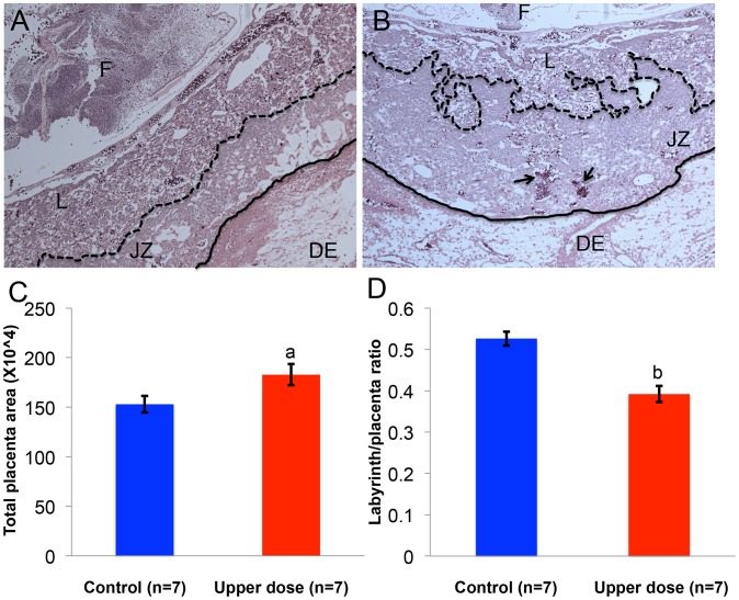
Figure 9. BPA exposure was associated with defective placental development.
Hematoxylin and eosin-stained histological sections of representative E12.5 conceptuses from (A) control and (B) upper dose BPA exposure groups are shown. F = fetus; DE = maternal decidua; L = labyrinth zone and JZ = junctional zone. Dotted line indicates the boundary between JZ and L, continuous line the boundary between JZ and DE, and arrows the accumulation of red blood cells. Total placenta area (C) and the ratio of area occupied by the labyrinth zone and total placenta area (D) were measured in both control (blue) and upper dose BPA exposed (red) placentas; a = P<0.05; b = P<0.001.

