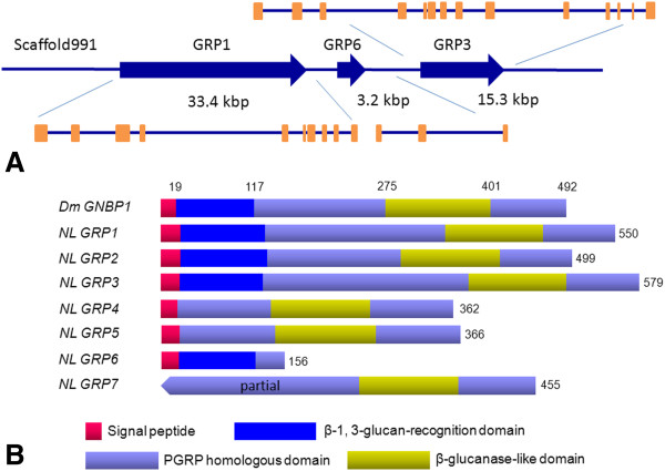
Figure 4.

(A) The gene structure prediction of N. lugens GRPs. The blue arrows indicate the transcription orientations and sizes of GRP1, GRP3 and GRP6 genes on scaffold991. The exons are shown with orange boxes. (B) The schematic representation of N. lugens GRPs. The deduced N. lugens GRP sequences were compared with D. melanogaster GNBP1 (CAJ18915). The putative signal peptide, N-terminal β-1, 3-glucan-recognition domain, PGRP homologous domain and C-terminal β-glucanase-like domain are shown in different color boxes. The number indicates the deduced amino acid residues.
