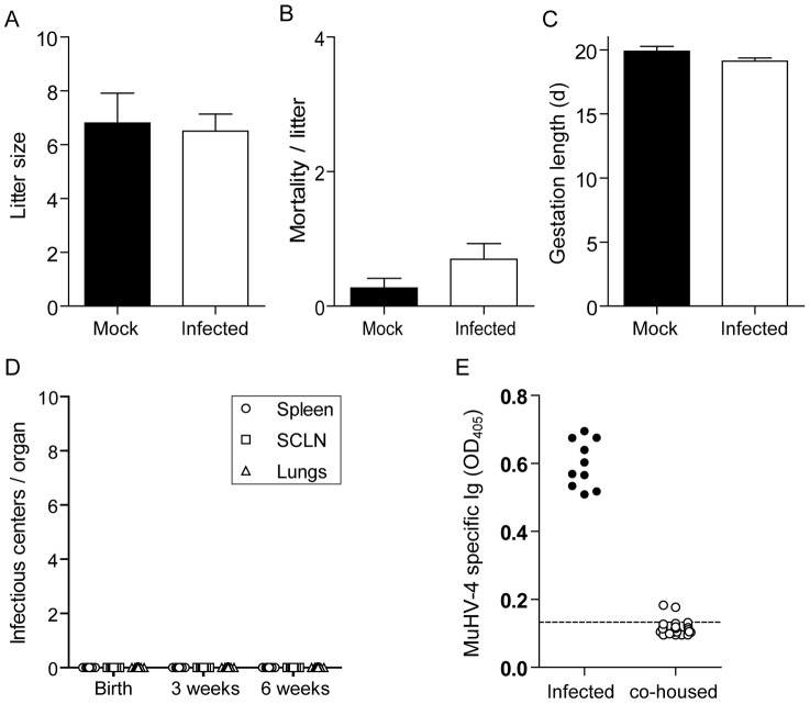
Figure 5. Vertical and non sexual transmissibility of MHV-68 from virus-excreting mice.
A–D. Female mice were infected intranasally (104 PFU) with WT luciferase+ MHV-68 under general anaesthesia, and then injected with luciferin and imaged every day. At the time of the first observation of genital signal, infected females were mated with uninfected males. Mock infected female mice were used as controls. Effect of MHV-68 infection on litter size (A), mortality/litter (B) and gestation length (C) was then monitored. The data presented are the average for 20 (infected) and 11 (Mock) pregnancies +/− standard error of the mean and were analyzed by 1way ANOVA and Bonferroni post-tests, no statistically significant difference was observed. Transmission to the progeny (n≥20 per group) was assessed by infectious center assays performed on isolated organs taken from newborn or at 3 or 6 weeks after birth (C). Data are plotted individually. E. Female mice (n = 10) were infected intranasally (104 PFU) with WT luciferase+ MHV-68 under general anaesthesia, and then injected with luciferin and imaged every day. At the time of the first observation of genital signal, infected females were co-housed with 3 uninfected females. Potential MHV-68 transmission was monitored 45 days later by detection of anti-MHV-68 specific antibodies. The dashed line indicates the mean value obtained with sera from 3 uninfected mice taken as controls.

