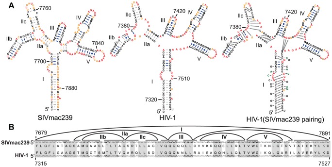
Figure 6. Codon alignment and predicted pairing partners in the RRE region of SIVmac239 and HIV-1NL4-3.
(A) Predicted structures within the RRE are shown for SIVmac239 (left) and HIV-1 (middle). Codons of stem I are in brackets with their corresponding amino acids labeled and numbered from the first codon of stem I. Blue brackets indicate an area of conserved pairing partners; green brackets indicate an area of shifted pairing partners in stem I. The HIV-1NL4-3 structure with forced SIVmac239 pairing is also shown with sequence variations that occur in SIVmac239 indicated while those that change the amino acid sequence are in green (right). Blue lines indicate base pairs that are exactly conserved between the two viruses. (B) The sequences of SIVmac239 (top) and HIV-1NL4-3 (bottom) aligned horizontally. Curved lines indicate base-pairing partners. Gray boxes indicate regions of amino acid alignment. Roman numerals indicate helices discussed in the text.

