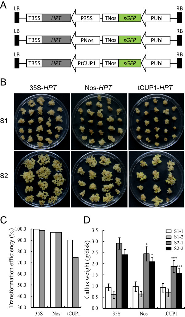
Figure 3.
The promoter activity of tCUP1 compared with 35S and Nos promoters. A. Schematic diagram showing the structure of the three HPT-GFP vectors. These vectors were constructed based on a modified pCAMBIA0380 vector by introducing the HPT gene driven by 35S, Nos and tCUP1 respectively. A Ubi promoter controlled GFP expression cassette was also introduced into each vector as a visual indicator for successful transformation. B. Growth of transformed calli 21 days after the onset of first round selection on 50 mg/ml Hyg (S1) and 14 days after the onset of second round selection on fresh selection medium with the same Hyg concentration (S2). Left panel, transformed with 35S controlled HPT-GFP vector; middle panel, transformed with Nos controlled HPT-GFP vector; right panel, transformed with tCUP1 controlled HPT-GFP vector. A representative dish of calli in each transformation is shown. C. Transformation efficiency of the three HPT-GFP vectors with HPT regulated by 35S, Nos and tCUP1 respectively. Transformation efficiency was calculated as a percentage from the numbers of GFP expressing calli and the numbers of primary calli infected 18 days after the onset of the first round of selection. Data is shown from two independent transformations (white and gray bars) with each vector. D. Hyg-tolerant growth of the calli transformed by the three HPT-GFP vectors with HPT regulated by 35S, Nos and tCUP1 respectively. Weight of calli per dish was measured at the beginning of the first (S1) and second (S2) rounds of selection. Data are shown as mean ± SD (n=6). The experiments were performed twice in two independent transformations (indicated as -1 and -2) with each vector. * and *** indicate a significant difference at P<0.05 and 0.001 respectively, in a Student t-test.

