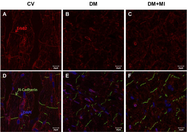
Figure 5.
Immunolocalization of ErbB2 receptor in failing diabetic post-MI rat heart. Cross-sections of rat heart tissue (A-F) were immunostained for ErbB2 (red), N-Cadherin (green), and DAPI (blue), visualized by confocal microscopy. Controls lacking primary anti-ErbB2 antibody showed minimal background staining (data not shown). Bar represents 20 μM. Immunolocalization images representative of 5–6 animals per group.

