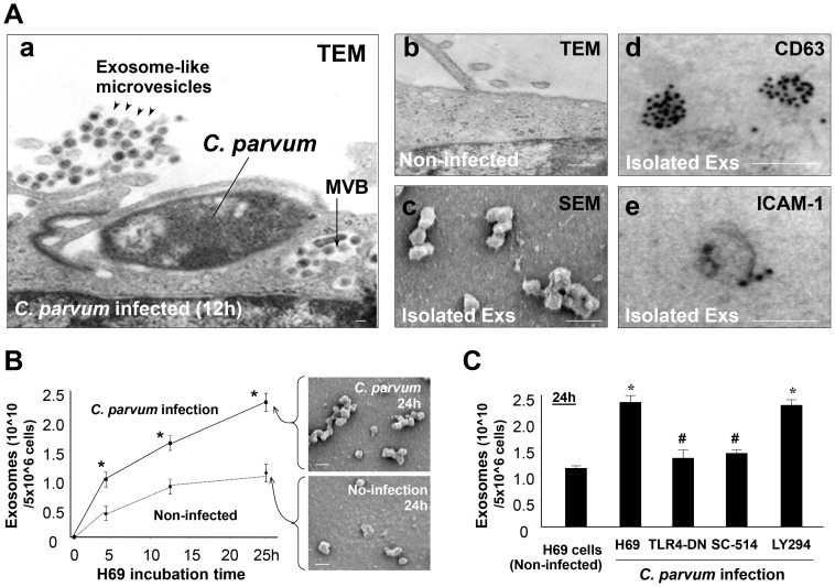
Figure 1. C. parvum infection increases luminal release of epithelial exosomes through activating the TLR4 signaling pathway.
(A) Infection stimulates luminal release of exosomes (Exs) from H69 monolayers, as assessed by electron microscopy (EM). Abundant exosome-like microvesicles were observed in the apical region of H69 monolayers following infection (arrowheads in a), with multi-vesicular bodies (MVBs) detected in the cytoplasm of infected cells (arrow in a). Only a few microvesicles were detected at the apical region of non-infected monolayers (b). Isolated apical microvesicles are 40–100 nm in size by scanning EM (c) and are positive by immunogold for exosome markers CD63 (d) and ICAM-1 (e). (B) A time-dependent apical exosome release was detected in infected H69 monolayers by nanosight tracking analysis and scanning EM. (C) Inhibition of TLR4 (by TLR4-DN transfection) and IKK2 (by SC-514), but not PI-3K (by LY294002), blocked C. parvum-induced exosome release. Scale bars = 100 nm; * p<0.05 ANOVA versus the non-infected controls; # p<0.05 ANOVA versus infected cells.

