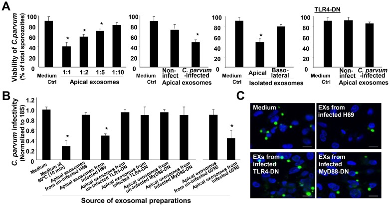
Figure 7. Apical exosomes released from C. parvum-infected biliary epithelium display anti-C. parvum activity.
(A) Effects of incubation with isolated exosomes from the biliary epithelium on C. parvum viability. Exosomes were isolated from the basolateral or apical supernatants of H69 monolayers of non-infected control, cells of C. parvum infection for 24 h, and cells stably expressing TLR4-DN after infection for 24 h. Freshly excysted C. parvum sporozoites were incubated with isolated exosomes (as indicated) for 2 h, and parasite viability was assessed by propidium iodide staining. (B) and (C) Effects of incubation with isolated apical exosomes from the biliary epithelium on the infectivity of C. parvum sporozoites. The same number of freshly excysted C. parvum sporozoites was incubated with exosomes isolated from H69 or 603B monolayers for 2 h and then added to H69 cells for 2 h. Parasite infection burden was measured by real-time PCR (B) and fluorescence microscopy (C). Pre-incubation of isolated exosomes abolished their inhibitory effects on C. parvum infectivity. C. parvum parasites were stained in green using a specific antibody and cell nuclei in blue by DAPI. Scale bars = 5 µm; *, p<0.05 ANOVA versus medium control.

