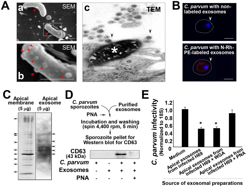
Figure 8. Gal/GalNAc-associated molecules mediate direct binding of exosomes to the C. parvum surface.
(A) Direct binding of apical exosomes to the C. parvum surface observed under EM. When sporozoites were added to H69 cells for 2 h, exosome-like microvesicles on their surface were observed, as evident by SEM (arrows in a and b; b shows a higher magnification of the boxed region in a). The TEM image shows a dying C. parvum parasite that was invading an epithelial cell with several exosomes binding to its surface (arrows in c). (B) Delivery of exosomal content to C. parvum sporozoites after incubation. Isolated exosomal vesicles were labeled with the N-Rh-PE dye, and after extensive washing, were incubated with C. parvum sporozoites for 2 h. These sporozoites showed positive labeling of the dye after incubation. Parasite nuclei were labeled in blue with DAPI (arrows). (C) Gal/GalNAc-associated molecules in H69 apical membrane fraction and exosomes released from H69 monolayers. Positive bands were detected in both H69 apical membrane fraction and released exosomes using SDS gel lectin-blotting with the Gal/GalNAc-specific lectin PNA. (D) Blotting of whole lysates of C. parvum sporozoites after pre-incubation with isolated exosomes revealed a positive reaction to exosome marker CD63. (E) PNA attenuates the inhibitory effects of exosomes on C. parvum viability. Pre-incubation of isolated exosomes abolished their inhibitory effects on C. parvum viability. Scale bars = 1 µm; *, p<0.05 ANOVA versus medium control.

