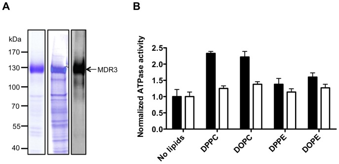
Figure 6. Characterization of purified human MDR3 in Fos-choline-16.

A Coomassie Brilliant Blue-stained SDS-PAGE and immunoblot using an anti-MDR3 antibody of purified MDR3 wild-type and the MDR3 EQ/EQ-mutant via TAP. Molecular weight markers are shown on the left. B Normalized ATPase activity of MDR3 wild-type (black) and of an ATPase deficient mutant (E558Q E1207Q, white) in FC-16 without and with different phospholipids. The ATPase activity of three independent MDR3 purifications was determined ± SD (n = 3).
