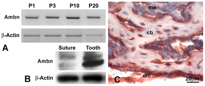
Figure 1. Ambn expression in cranial bone and dura mater.

(A) RT-PCR analysis of Ambn mRNA expression in the posterior frontal suture region from 1, 3, 10, and 20 days postnatal WT mice. The β-Actin gene was used as internal control. (B) Western blot analysis of AMBN protein expression in postnatal day 3 posterior-frontal (PF) suture tissues and teeth. Two bands at 55 and 50 kDa were recognized. β-Actin was used as loading reference. (C) Immunostained sections of cranial bone and dura mater from 3 day postnatal wildtype (WT) mice demonstrated Ambn protein localization in calvarial bone (cb), dura mater (dm), and condensed mesenchymal cells (me). Note the high levels of Ambn protein in the dura mater and in calvarial osteoblasts. Bar = 20 µm.
