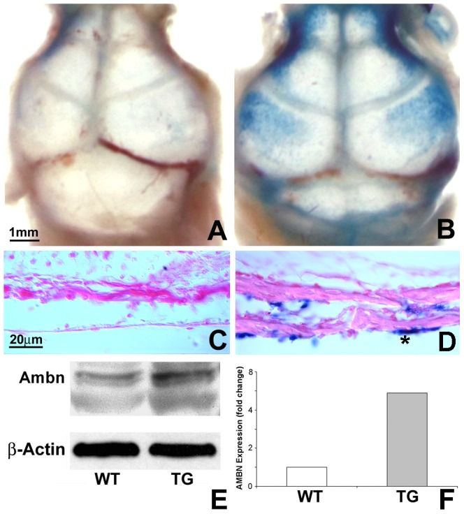
Figure 2. LacZ and Ambn transgene expression in cranial bone and sutures.

(A and B) Whole mount β-gal staining of wildtype (WT) and K14-LacZ overexpressing mouse skulls. The β-gal blue signal was localized in cranial bones and sutures of the Ambn overexpressor. (C and D) Histological sections of β-gal stained and Eosin counterstained skulls were prepared from the coronal plane of the calvaria in the posterior-frontal region. Note the β-gal positive calvarial osteoblasts in the Ambn transgenic mouse sections. No blue staining of β-gal was detected in the WT skull section. Bar for A and B = 1 mm, C and D = 20 µm. (E) Western blot analysis of protein extracts from WT and Ambn transgenic calvarial bone and sutures at age postnatal day 3. The Ambn protein level in the transgenic mouse (right lane, upper panel) was 5-fold higher than that in the control mouse (left lane, upper panel) after normalization with β-actin (lower panel).
