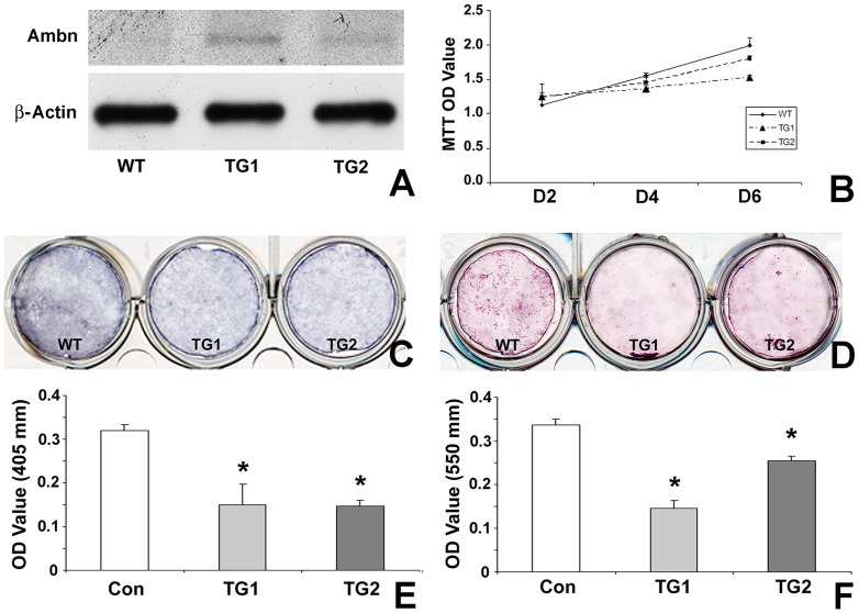
Figure 6. Effect of Ambn on proliferation and differentiation of suture mesenchymal cells.
In this study, mesenchymal cells from wildtype (WT) mice and Ambn overexpressor (TG) sutures were cultured in osteogenic induction medium. (A) Ambn protein expression in suture-derived cells. β-Actin was used as loading control. (B) MTT assay to compare cell proliferation in WT and TG mesenchymal suture cells. The measured absorbance (mean+/− SD) is proportionate to the number of living cells. (C) Alkaline phosphotase activity staining at day 6 of culture identified differentiated cells positive for alkaline phosphatase with blue staining. (D) Alizarin red staining for mineral nodule formation. Cells were cultured for 21 days in osteogenic induction medium. Mineral nodules were stained in red. All experiments were carried out in triplicate and repeated three times. * represents significant difference, p<0.05 (Kruskall-Wallis one-way analysis).

