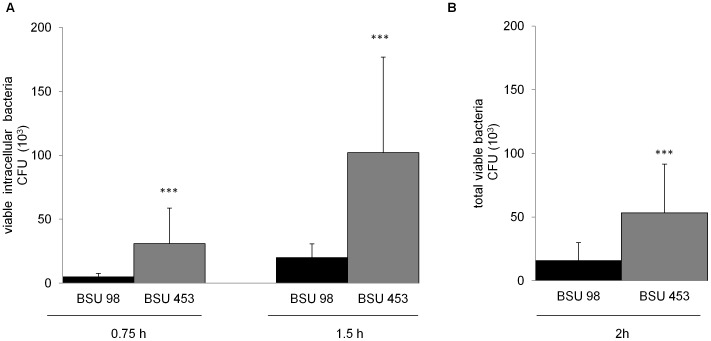
Figure 1. Survival of S. agalactiae and β-hemolysin expression in professional phagocytes.
The monocyte-derived macrophage cell line THP-1 or freshly isolated granulocytes were infected with hemolytic (BSU 98) and nonhemolytic (BSU 453) bacteria at a MOI of 1∶1 for indicated time points. A) Intracellular bacteria were quantified after killing the extracellular bacteria using Penicillin (1 µg/ml) and Gentamicin (100 µg/ml) for additional 1 h. B) Total viable bacteria after incubation with granulocytes without killing of extracellular bacteria. Data shown are the mean ± SD of six independent experiments. Data is considered extremely significant for p values <0.001 (***).

