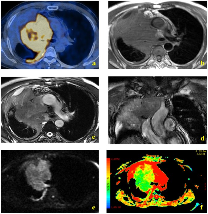
Figure 2. MR and PET/CT images of a 58-year-old man with small cell lung carcinoma.
PET-CT indicated greater uptake of FDG by tumor than atelectasis (a). T1W MR imaging showed a soft-tissue shadow in the right upper lobe, but similar signal intensities of the central lung carcinoma and the distal atelectasis were noted (b). FRFSE T2W axial and coronal images indicated hypointensity of the tumor mass relative to the atelectasis (c and d, respectively). DW images obtained with a diffusion gradient of 500 s/mm2 allowed easy differentiation of tumor and atelectasis (e and f).

