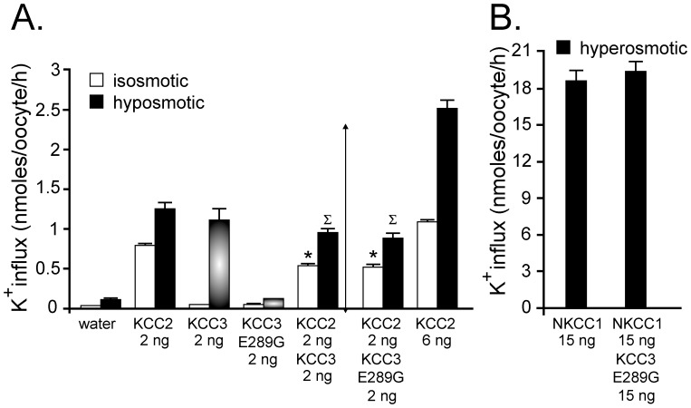
Figure 3. Evidence for dominant-negative effect of KCC3 on KCC2 function.
A) Xenopus laevis oocytes were injected with KCC2 cRNA in the presence or absence of wild-type KCC3 or KCC3-E289G cRNAs. Three days following injection, K+ influx was measured under isosmotic conditions where only KCC2 function is active, or under hyposmotic conditions where both KCC2 and KCC3 function are stimulated. Bars represent mean ± S.E.M. (n = 20–25 oocytes). Flux is expressed in nmoles K+/oocyte/h. (*) P<0.01 (ANOVA) compared with KCC2 alone under isosmotic condition, (∑) P<0.01 (ANOVA) compared with KCC2 alone under hyposmotic solution. Arrow indicates the anticipated flux mediated by the combined activity of KCC2 and KCC3. This is a representative experiment. Each condition (bar) was reproduced multiple times. B) K+ influx was measured under hyperosmotic conditions in oocytes injected with NKCC1 cRNA in the presence or absence of KCC3-E289G cRNA. Bars represent mean ± S.E.M. (n = 20–25 oocytes). Flux is also expressed in nmoles K+/oocyte/h.

