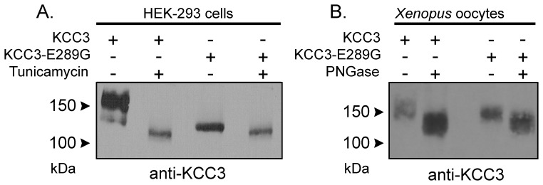
Figure 7. N-Glycolsylation deficiency of the KCC3-E289G mutant.
A) Western blot analysis of wild-type KCC3 and KCC3-E289G mutant in HEK 293FT cells treated with tunicamycin (10 µg/ml for 18 h). B) Western blot analysis of wild-type KCC3 and KCC3-E289G mutant proteins isolated from Xenopus laevis oocytes and treated with PNGase (0.25U, 12 h at 37°C). The membranes were exposed to a rabbit polyclonal anti-KCC3 antibody. The experiment was repeated once with identical data.

