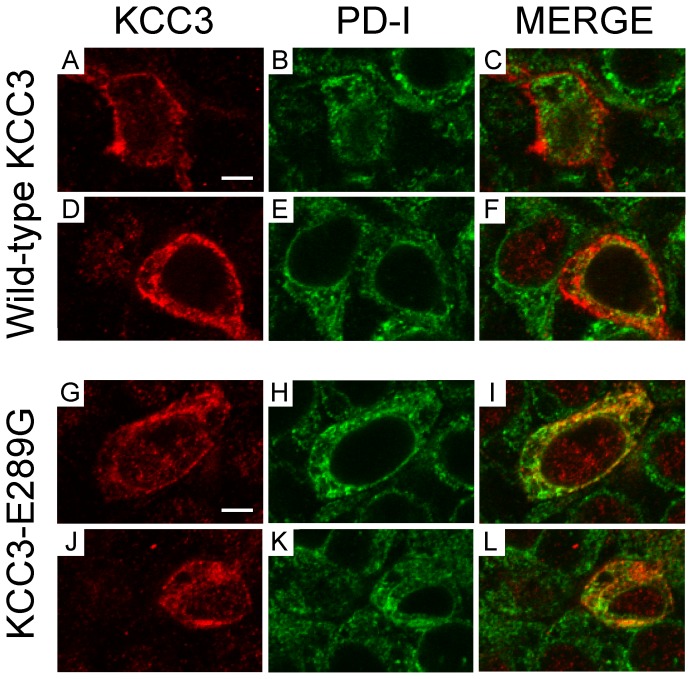
Figure 8. Evidence for KCC3-E289G localizing in the endoplasmic reticulum.
HEK 293FT cells were transfected with wild-type KCC3 (A–F) or KCC3-E289G mutant (G–L). Two days post- transfection, the cells were fixed with paraformaldehyde, treated with saponin, and exposed to rabbit polyclonal anti-KCC3 and mouse monoclonal anti-PDI antibodies followed by cy3-conjugated anti-rabbit and Alexa Fluor–conjugated goat anti-mouse antibodies. Focal plane images of KCC3 signal (A, D, J, G), ER marker signal (B, E, H, K), and combined signals (C, F, I, L). Bar = 5 µm.

