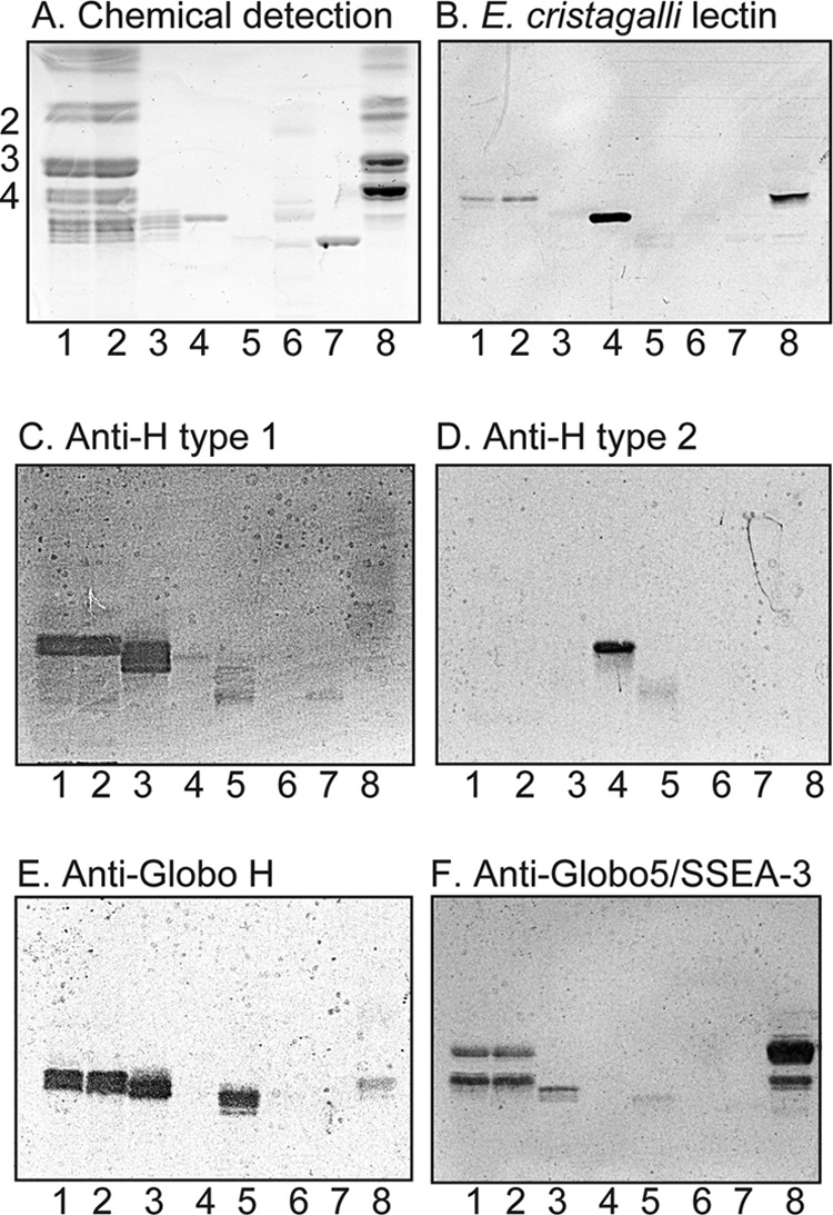
FIGURE 1.

Binding of E. cristagalli lectin and monoclonal antibodies to non-acid glycosphingolipids of human embryonic stem cells. A–F, thin-layer chromatogram after detection with anisaldehyde (A) and autoradiograms obtained by binding of E. cristagalli lectin (B), the monoclonal anti-H type 1 antibody 17-206 (C), the monoclonal anti-H type 2 antibody 92FR-A2 (D), the monoclonal anti-Globo H antibody MBr1 (E), and the monoclonal anti-Globo5/SSEA-3 antibody MC-813-70 (F). Lane 1, non-acid glycosphingolipids of human embryonic stem cell line SA121 (40 μg); lane 2, non-acid glycosphingolipids of human embryonic stem cell line SA181 (40 μg); lane 3, reference H type 1 pentaglycosylceramide (Fucα2Galβ3GlcNAcβ3Galβ4Glcβ1Cer) (4 μg); lane 4, reference H type 2 pentaglycosylceramide (Fucα2Galβ4GlcNAcβ3Galβ4Glcβ1Cer) (4 μg); lane 5, reference Globo H hexaglycosylceramide (Fucα2Galβ3GalNAcβ3Galα4Galβ4Glcβ1Cer) (4 μg); lane 6, non-acid glycosphingolipids of mouse small intestine (10 μg); lane 7, reference B type 2 hexaglycosylceramide (Galα3(Fucα2)Galβ4GlcNAcβ3Galβ4Glcβ1Cer) (4 μg); lane 8, non-acid glycosphingolipids of human kidney (blood group A) (20 μg). The numbers to the left of the chromatogram in A denote the approximate number of carbohydrate residues in the bands. The chromatograms were eluted with chloroform/methanol/water (60:35:8, v/v/v), and the binding assays were done as described under “Experimental Procedures.” Autoradiography was for 12 h.
