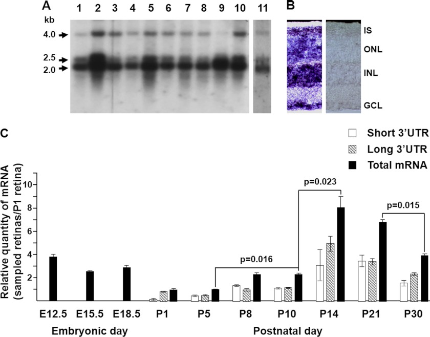
FIGURE 1.
Rhbdd2 mRNA expression in mouse tissues and its quantification during retinal development. A, a mouse multitissue Northern blot of mRNAs from heart (lane 1), brain (lane 2), liver (lane 3), spleen (lane 4), kidney (lane 5), embryo (lane 6), lung (lane 7), thymus (lane 8), testis (lane 9), and ovary (lane 10) and a separate blot containing mouse retinal mRNA (lane 11) were probed with a 32P-labeled Rhbdd2 cDNA. Arrows indicate the sizes of the three detected transcripts (2.0, 2.5, and 4 kb), respectively. B, in situ hybridization of Rhbdd2 mRNAs in mouse retina with antisense (left panel) and sense (right panel) DIG-labeled riboprobes. The Rhbdd2 mRNA localized to all the nuclear layers; no signal was detected with the sense probe. C, quantitative RT-PCR amplification of mRNAs from mouse embryonic eyes and postnatal retinas at different times of development. Relative expression of Rhbdd2 is normalized to the β-actin mRNA level. Error bars indicate S.E. IS, inner segment; ONL, outer nuclear layer; INL, inner nuclear layer; GCL, ganglion cell layer.

