FIGURE 3.
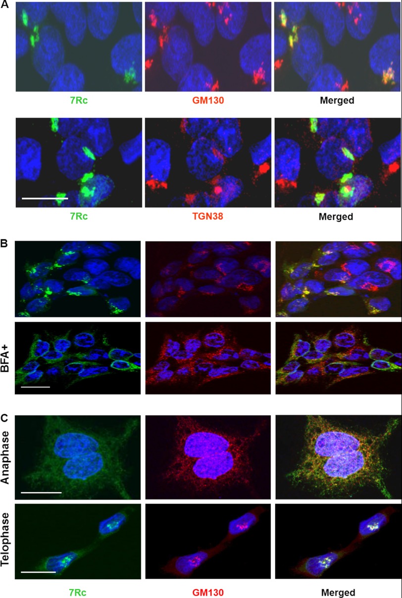
Golgi localization of RHBDD2 in transfected HEK293 cells. A, immunocytochemical detection of RHBDD2 and GM130 (top panel) or RHBDD2 and TGN38 (bottom panel) proteins after transient transfection of the Rhbdd2 expression vector into HEK293 cells. The obtained images for each staining were merged. Scale bar, 15 μm. B, Rhbdd2-transfected cells with or without 50 μm BFA were incubated for 30 min at 37 °C before fixation. Cells were stained with 7Rc or anti-GM130 antibody. RHBDD2 co-localization with GM130 is sensitive to the exposure of cells to BFA. C, RHBDD2 and GM130 co-localize during mitosis. RHBDD2-expressing cells were fixed, permeabilized, and double stained with the same antibodies during anaphase and telophase. Scale bars for B and C, 10 μm.

