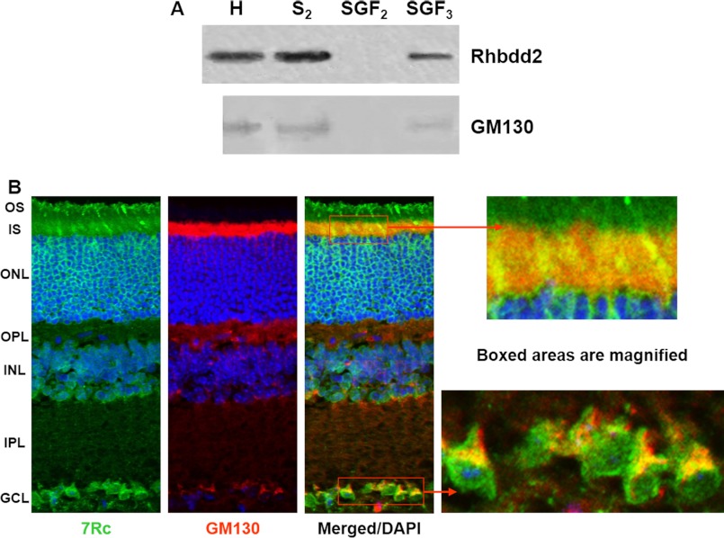
FIGURE 6.
Fractionation of mouse retinal protein extract and co-localization of RHBDD2 with a Golgi marker. A, the postnuclear supernatants were fractionated on discontinuous sucrose gradients. Western blots incubated with 7Rc and GM130 antibody show that the S2 fraction contains a substantial amount of RHBDD2 protein; upon recentrifugation of S2, a significant amount of RHBDD2 is found in the stacked Golgi fraction 3 (SGF3). H, homogenate. B, confocal microscopic images of adult mouse retinal sections stained with 7Rc, GM130 antibody, and DAPI. The merged image illustrates co-localization of these two proteins. The far right panel shows the magnified boxed areas. OS, outer segment; IS, inner segment; ONL, outer nuclear layer; OPL, outer plexiform layer; INL, inner nuclear layer; IPL, inner plexiform layer; GCL, ganglion cell layer.

