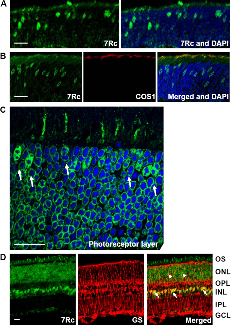
FIGURE 8.
Detection of RHBDD2 expression in young and adult mouse retina sections. A, section of a 12-day-old mouse retina stained with 7Rc-FITC and DAPI, which was used to stain the nuclei. The photoreceptor layer shows strongly stained cone cell nuclei migrating toward the outer edge of ONL and short outer segments. B, a 12-day-old mouse retinal section triple stained with 7Rc tagged with FITC, the cone marker COS1 tagged with Alexa Fluor 647, and DAPI. The merged image shows overlapping RHBDD2 and COS1 staining. C, a magnified image of the ONL from an adult retinal section immunolabeled with 7Rc. The arrows point to the larger, oval nuclei of cones that appear to contain several clumps of irregularly shaped heterochromatin. D, images of an adult retina showing RHBDD2 localization (green), glutamine synthetase (GS) localization (red), and their co-localization in the somas of many Müller glial cells (arrows) and some of their processes (arrowheads). Scale bars, 20 μm. INL, inner nuclear layer; OPL, outer plexiform layer; IPL, inner plexiform layer.

