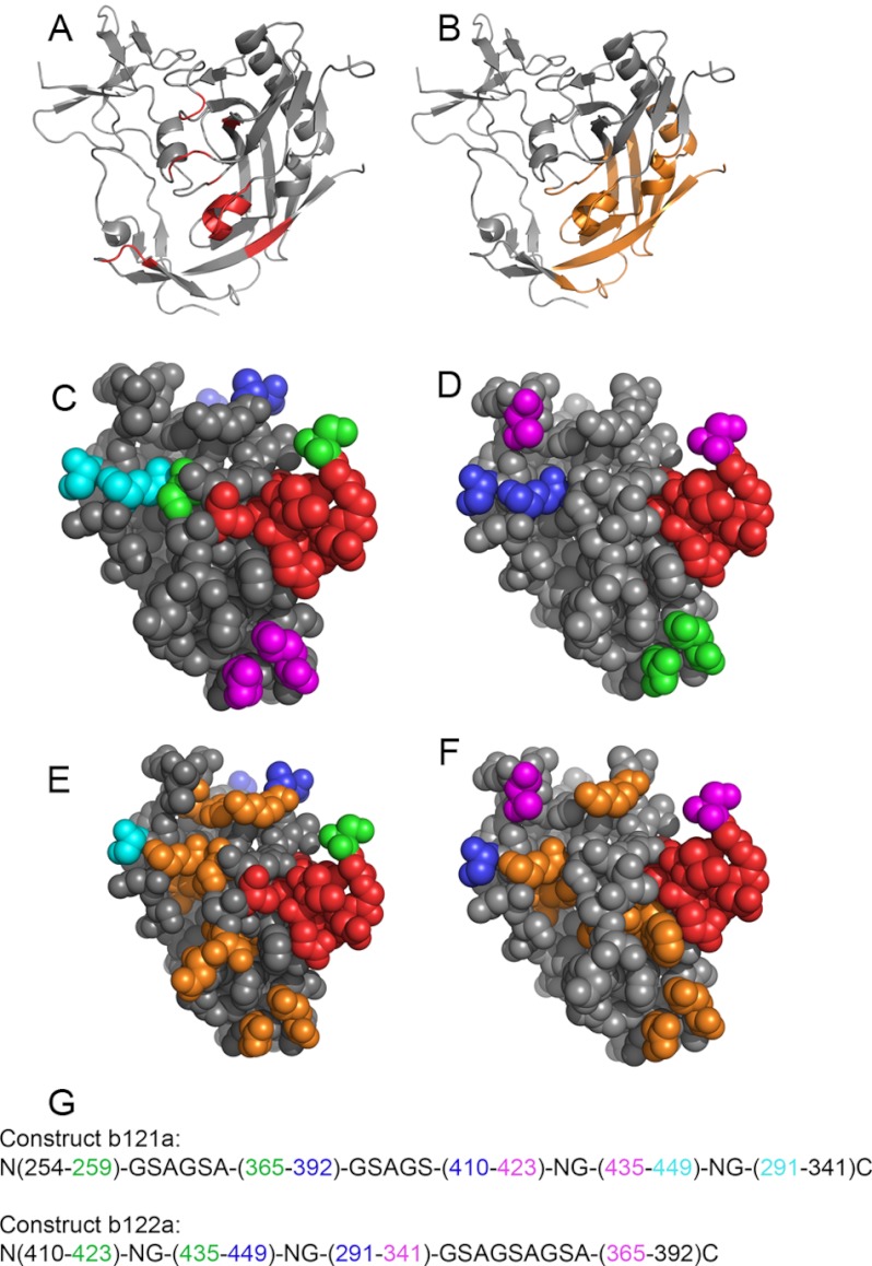
FIGURE 1.
Structure of core gp120 when complexed to the broadly neutralizing antibody b12. The coordinates are from Protein Data Bank entry 2NY7. A, residues involved in binding to b12 are shown in red; B, regions in gp120 included in b121a and b122a are shown in orange and include ∼70% of the binding site. Space-filled structures of the regions included in b121a and b122a are shown in C and D, respectively. The b12 binding site is colored in red. Residues connected by linkers are in the same color. Similar space-filled models of b121a and b122a are shown in E and F, where shown in orange are exposed hydrophobic residues that have been mutated to suitable polar residues based on Rosetta calculations and visual inspection. G, connectivities for b121a (top) and b122a (bottom). Colors are identical to those used in C and D, respectively.

