FIGURE 5.
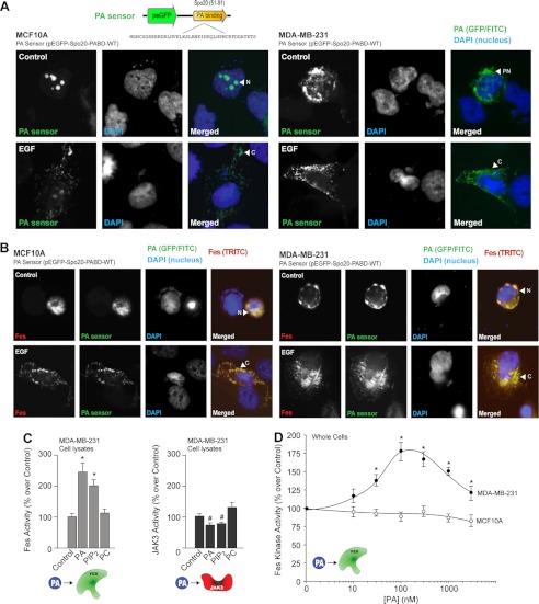
Interplay between PA and Fes and JAK3 kinases. A, MCF10A and MDA-MB-231 cells were plated onto glass coverslips and then transfected with 1 μg of Fes plasmid in combination with 1 μg of the PA sensor (EGFP-based) plasmid. Green denotes the EGFP-tagged PA sensor. Red denotes the TRITC-tagged Fes. Blue denotes DAPI staining of the nucleus. Shown is a staining of the PA sensor (FITC) and nuclei (DAPI) only in cells in the absence (upper row) or presence (lower row) of EGF. B, similar to above, this time in cells co-expressing the PA sensor and Fes (TRITC). C, effect of 100 nm lipids (PA, PIP2, or PC) on recombinant JAK3 and Fes activities in vitro. D, differential effect of PA (1,2-dioleoyl-sn-glycero-3-phosphate, PA in a di-oleoyl or membrane-soluble form) on endogenous Fes activity.

