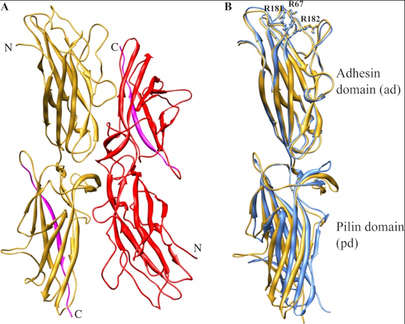
FIGURE 4.
A, two dscCfaE G168D molecules (yellow and red) are present in one asymmetric unit in the crystal structure. The donor strands are highlighted in cyan. B, superposition of chain A from both the dscCfaE (blue) and dscCfaE G168D (yellow) structures. The side chains of three arginines (Arg-67, Arg-181, and Arg-182) are shown in ball and stick.

