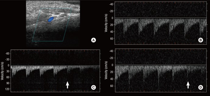
Fig. 2.
Doppler sonogram of the pulsating flow in the mastoid emissary vein (MEV), correlating to the pulsatile tinnitus. Doppler sonography identified the location of the outer foramen of the MEV and the pulsating venous blood flow draining into the sigmoid sinus (A, B). The patient recognized reduction of the pulsatile tinnitus synchronously with the decrease of the flow-signal by compression of the left jugular vein (C) and the Valsalva maneuver (D). Arrows: starting points of compression of the left jugular vein and the Valsalva maneuver, respectively.

