
Figure 2.
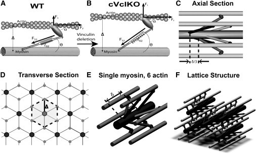
Micromechanical model for sarcomere lattice spacing and cross-bridge architecture. (A) Individual cross-bridge model consisting of a myosin stalk based on the geometry by Schoenberg (19). (B) As the lattice spacing (Δ) increases with Vcl deletion (transition from panels A to B), binding angles (θ) change and FS2 increases, and attachment angle α remains constant. The result is an increase in the transverse force, but little change in the fiber force (Ft = transverse force, Ff = fiber force, FS2 = force along S2 segment). (C and D) The unit cell of the lattice structure in the axial (C) and transverse (D) directions is marked (dotted outline, δ = 3 pairs of myosin heads spacing). (E) A single myosin filament is surrounded by six actin filaments. (F) Overall lattice structure. Details of this myofilament lattice model can be found in the Supporting Material.