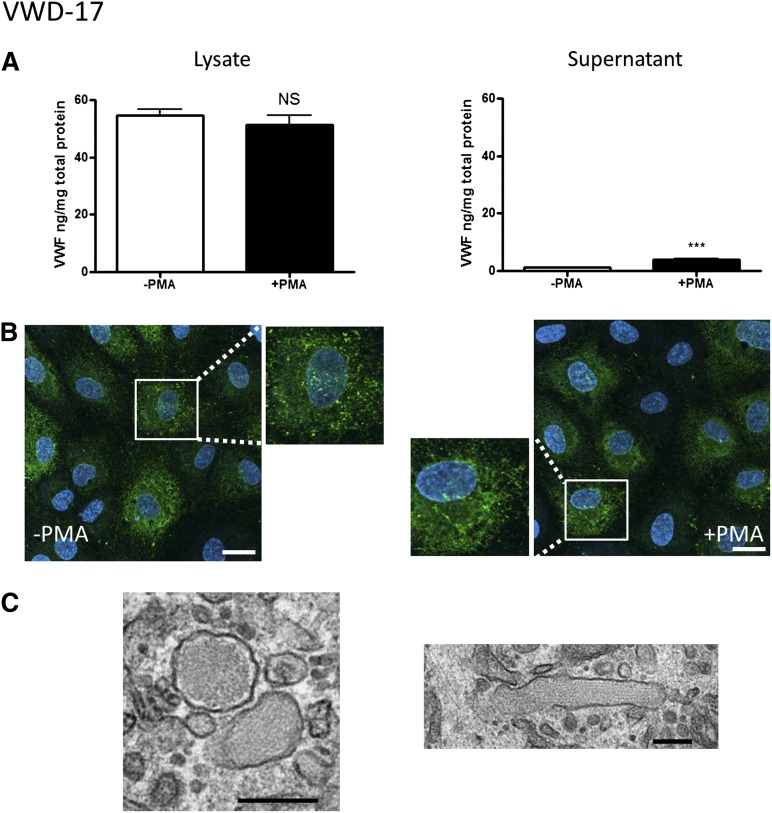
Figure 7.
BOEC characterization: type 2M VWD. (A-C) Data from patient VWD-17. (A) VWF was measured by ELISA in the total cell lysate (left) and cell culture supernatant (right) from BOECs in the absence or presence of PMA stimulation (mean ± SEM from 3 replicates). (B) VWF expression from control and PMA-stimulated BOECs was visualized by confocal IF microscopy (scale bars represent 20 µm). Boxes show 2× magnified image. (C) EM analysis of BOECs (scale bars represent 200 nm) shows the presence of rounded WPBs (left), plus a rare elongated WPB (right). ***P < .001.

