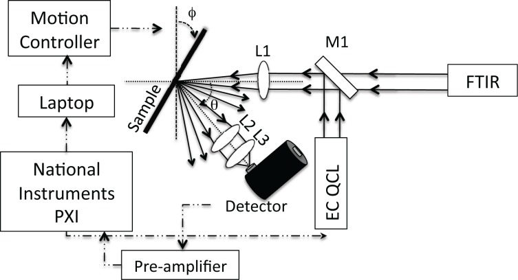
Fig. 1.
Schematic drawing of the experimental setup. A mirror (M1) was attached to a flip mount to allow for either source, the FTIR or the EC-QCL, to be used without realignment of the optical beam path. A lens (L1) was used to focus the collimated light onto the sample. Two lenses (L2 and L3) were used to collect the scattered light from the skin onto the detector.

