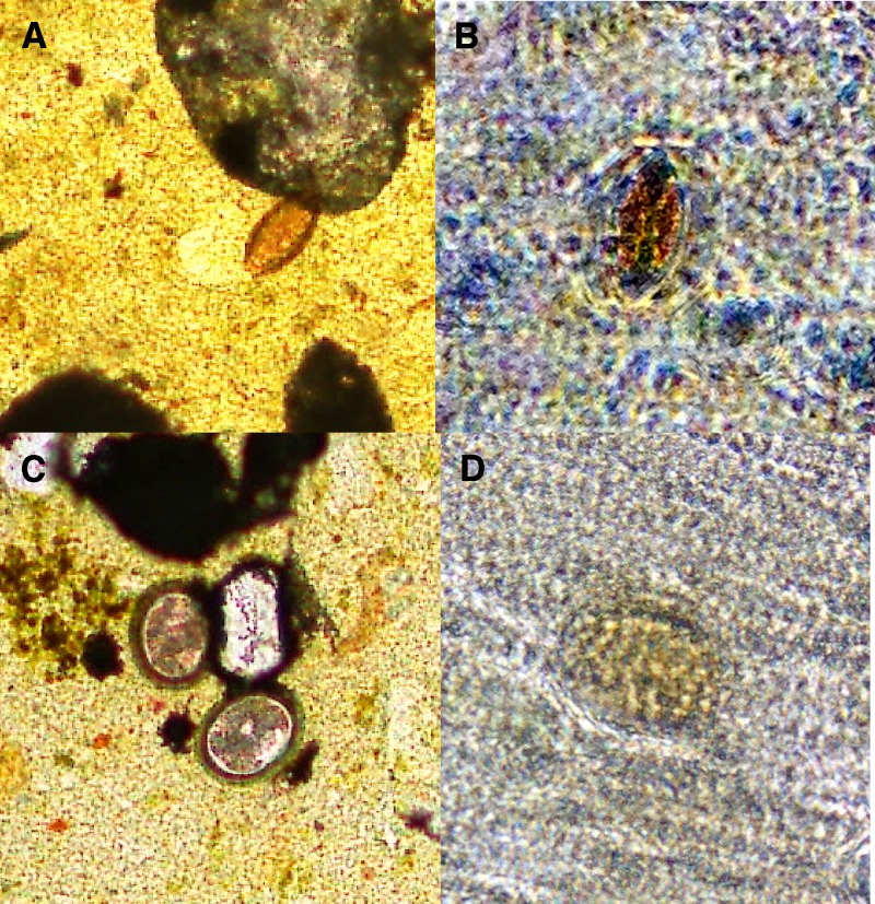
Figure 2.
Soil-transmitted helminth eggs visualized by conventional and mobile phone microscopy. T. trichiura is shown at 40× magnification by conventional microscopy in A and mobile phone microscopy in B. A. lumbricoides is shown at 40× magnification by conventional microscopy in C and mobile phone microscopy in D.

