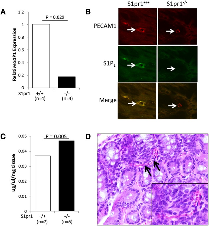
Fig. 2.
Genetic deletion of S1pr1 results in colonic vascular fragility. A: Colonic tissue was harvested from S1pr1+/+ and S1pr1−/− mice, and S1pr1 expression was determined by qRT-PCR. A statistically significant reduction in S1pr1 expression [median (range)] was found in S1pr1−/− [0.2 (0.1–0.3)] compared with S1pr1+/+ [1.0 (0.9–1.1)] mice. B: Tissues from mice described in panel A were examined by coimmunofluorescence for the expression of S1P1 and PECAM1 (63× objective). Note the loss of S1P1 expression on PECAM1-expressing cells in the colons of S1pr1−/− mice. Images were cropped and magnified to enhance visualization of individual blood vessels. C: Evans Blue dye was injected into the tail veins of S1pr1+/+ and S1pr1−/− mice, and the extravasation of dye from colonic tissue was determined, as described in Experimental Procedures. A statistically significant increase in vascular permeability [median (range)] was found in colons from S1pr1−/− [0.047 (0.040–0.051)] compared with S1pr1+/+ [0.037 (0.032–0.040)] mice. D: A representative photomicrograph of a colon from an S1pr1−/− mouse showing red blood cells (arrows) in the colonic mucosa that are not contained within a blood vessel (400×). The inset shows a cropped and magnified version of this image to enhance visualization of red blood cells.

