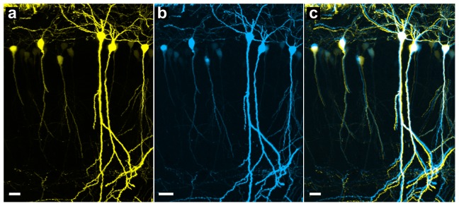
Figure 2. Morphology of pyramidal neurons (hippocampus) is well preserved after being embedded in optimized GMA resin.

A brain section from a Thy1-eYFP-H mouse imaged with two-photon microscopy before (a) and after embedding (b). (c) Merged images from (a) and (b). Scale bar, 20 µm. Each image is a max-projection of the image stacks of the brain slice (thickness: 60 µm).
