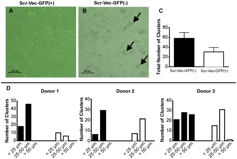
Figure 1. TurboGFP impairs T cell clustering after CD3/CD28 activation.
T cells were transfected with either the Scr-Vec-GFP(+) or the Scr-Vec-GFP(−) vectors, then activated by plate-bound anti-CD3 and soluble CD28 for 20 hours. A) GFP-expressing T cells exibit impaired clustering after CD3/CD28 activation. B) Normal cluster formation of CD3/CD28-activated T cells in sample transfected with the Scr-Vec-GFP(−) are indicated by black arrows. A representation of 3 independent experiments is shown. C & D) Activated cell clusters were quantified within one representative field of view at 100x for the Scr-Vec-GFP(−) vector and the Scr-Vec-GFP(+) vector transfections to obtain the total number of clusters (C), or the numbers of clusters by size range (<25 μm, 25–50 μm, and >50 μm) (D) where the black bars indicate the Scr-Vec-GFP(−) transfections and the white bars indicate the Scr-Vec-GFP(+) transfections. Mean and SEM are shown.

