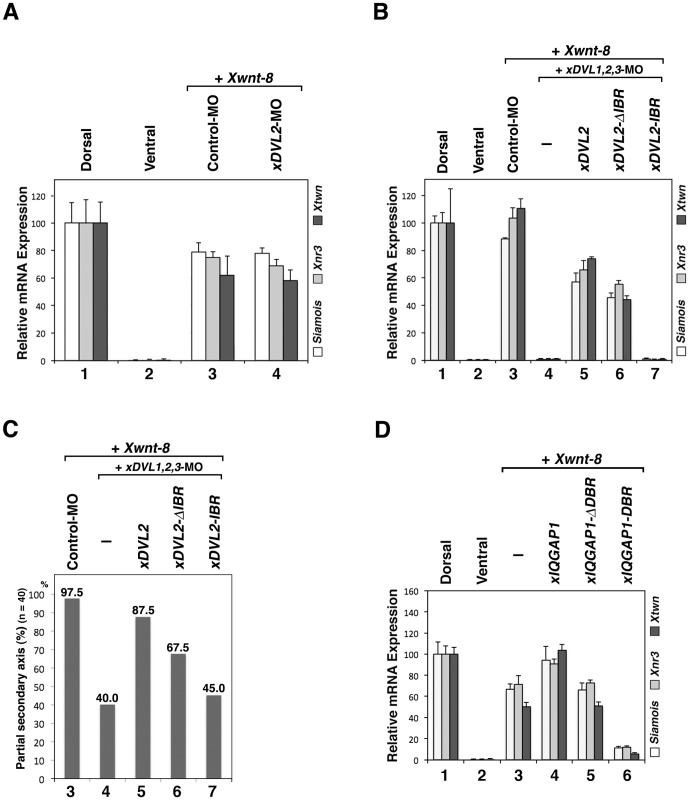
Figure 5. The role of the xIQGAP1-binding region of xDVL2 in canonical Wnt signaling during early embryogenesis.
(A) Quantitative RT-PCR analysis of early dorsal Wnt target genes (n = 3). Control-MO (15 ng) or xDVL2-MO (15 ng) was ventrally co-injected with Xwnt-8 (0.5 pg) mRNA. RNAs from dissected ventral sectors of injected embryos were extracted at stage 10. The following procedure is indicated in Figure 4A. Error bars represent standard deviation of the mean in three experiments. Statistical significance was determined by Student's t-test for each marker gene. The highest P values in three marker genes were chosen as a representative, as follows: P>0.1 [between lane 3 and lane 4]. (B) Quantitative RT-PCR analysis of early dorsal Wnt target genes (n = 3). xDVL1-MO (10 ng), xDVL2-MO (10 ng), xDVL3-MO (10 ng) and Xwnt-8 (0.5 pg) mRNA were ventrally co-injected with xDVL2 constructs: xDVL2 (25 pg), xDVL2-ΔIBR (25 pg), xDVL2-IBR (25 pg) mRNA. RNAs from dissected ventral sectors of injected embryos were extracted at stage 10. The following procedure is indicated in Figure 4A. P<0.05 [between lane 3 and lane 4], P<0.05 [between lane 4 and lane 5], P<0.1 [between lane 5 and lane 6], P<0.05 [between lane 5 and lane 7]. (C) The ratio of injected embryos that exhibited a partial secondary axis. The numbered lanes indicate the injected mRNAs and MOs consistent with the numbering in Figure B. (D) Quantitative RT-PCR analysis of early dorsal Wnt target genes (n = 3). Xwnt-8 (0.5 pg) mRNA was ventrally co-injected with xIQGAP1 constructs: xIQGAP1 (400 pg), xIQGAP1-ΔDBR (1 ng), xIQGAP1-DBR (1 ng) mRNA. The following procedure is indicated in Figure 4A. P<0.01 [between lane 3 and lane 4], P>0.1 [between lane 3 and lane 5], P<0.05 [between lane 3 and lane 6].

