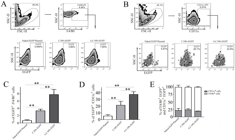
Figure 5. A.C.NPs loaded with DNA pass through the acidic gastric barrier and are taken up by macrophages and dendritic cells in the intestinal Peyer’s patches.
Naked EGFP DNA plasmids, C.NPs-EGFP, and A.C.NPs-EGFP were separately given to BALB/c mice (n = 5) via intragastric gavage at a daily dose equivalent to 30 µg plasmid DNA per mouse for three consecutive days. Peyer’s patches were isolated and analyzed by flow cytometry. PE-conjugated F4/80 and APC-conjugated CD11c antibodies were used to stain (A) the macrophages and (B) dendritic cells, respectively. Histograms of the percentage of EGFP-positive (C) macrophages and (D) dendritic cells. (E) Histograms of the ratio of F4/80- or CD11c- positive cells to total EGFP-positive cells. Data are presented as mean ± SD of three independent experiments (**P<0.01; n = 5).

