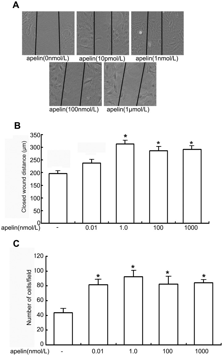
Figure 5. Migrating and chemotactic effects of apelin on glomerular endothelial cells.
A: The scratch-wound assay for glomerular endothelial cells was performed in the presence of apelin. The injury was performed by scraping the monolayer (the denuded area is between the panel lines). After 24 h of incubation in the presence or absence of apelin, cell migration into the wound edge was observed by light microscopy. The distance migrated to the denuded area is shown in graph. B: The decreased distance to the denuded area was significantly enhanced by apelin-13 at the concentration of 1 nmol/L compared to that in the control group (apelin at a concentration of 0 nmol/L). The data are expressed as the means±SD (n = 6, *p<0.05 vs. control group). C. Chemotactic effect of apelin on glomerular endothelial cells. The data are expressed as the means±SD (n = 6, *p<0.05 vs. control group).

