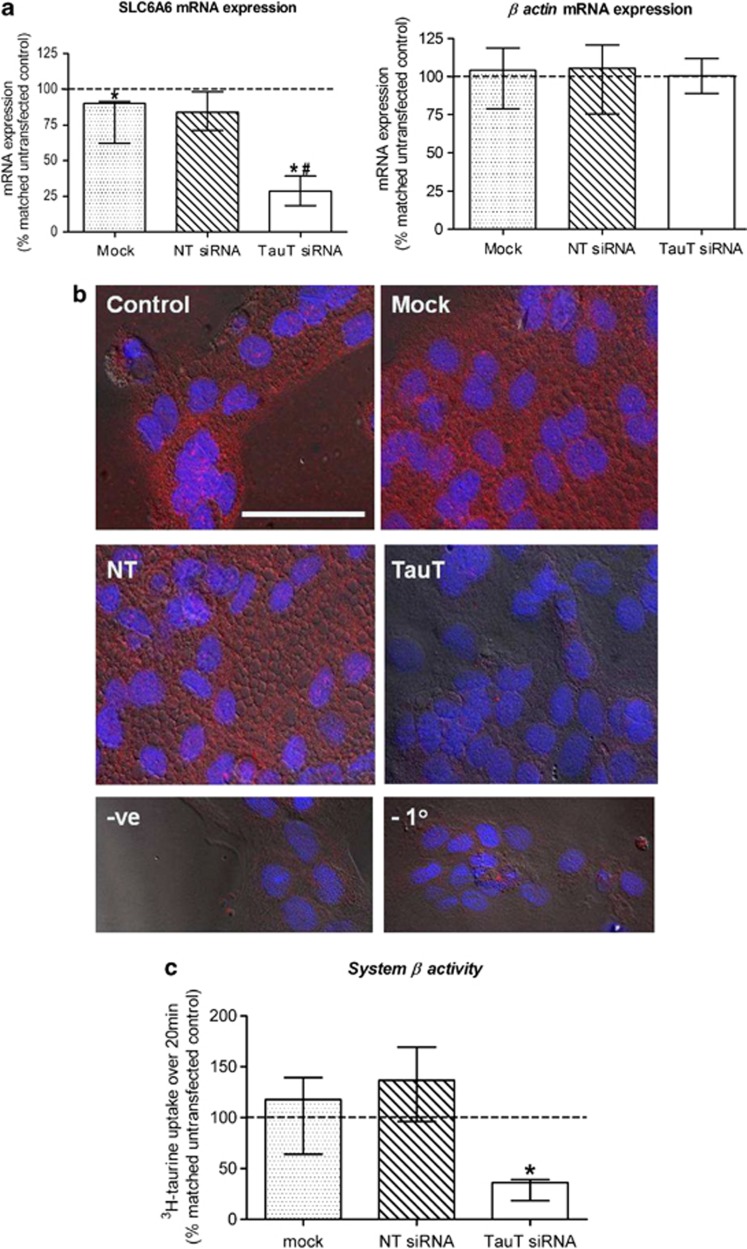
Figure 1.
Confirmation of TauT knockdown in 66-h cytotrophoblast cells. (a) SLC6A6 and β actin mRNA expression. (b) Immunofluorescent detection of TauT protein (red) in cells counterstained with DAPI (blue). Scale bar represents 50 μM and refers to all images. A lack of fluorescence following pre-absorption of primary antibody with a 10 × excess of antigenic peptide (−ve) or omission of the primary antibody (−1°) confirmed specificity. (c) TauT activity. All observations were made 48 h post transfection. Key to labelling: control=untransfected, mock=transfection reagent only, NT=non-targeting siRNA, TauT=SLC6A6-specfic siRNA. Error bars represent median±interquartile range, n=5. *P<0.05 versus 100% (i.e., matched untransfected control), Wilcoxon-signed Rank test. #P<0.05 versus mock and NT siRNA, Kruskal–Wallis with Dunn's multiple comparison test

