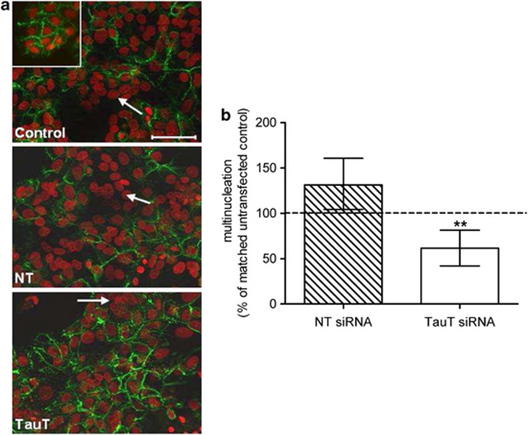
Figure 3.
TauT knockdown impairs cytotrophoblast morphological differentiation in vitro. (a) Immunofluorescent detection of desmosomes (green) in 18 h (inset) and 66 h cytotrophoblast cells counterstained with propidium iodide (red). The white arrows indicate multinucleated cells (defined as ≥3 nuclei within desmosomal boundaries). (b) Percentage of multinucleated cells at 66 h following transfection, relative to matched control cells (n=7, error bars represent median±interquartile range). **P<0.01 versus 100% Wilcoxon-signed Rank test. NT=non-targeting siRNA, TauT=SLC6A6-specfic siRNA

