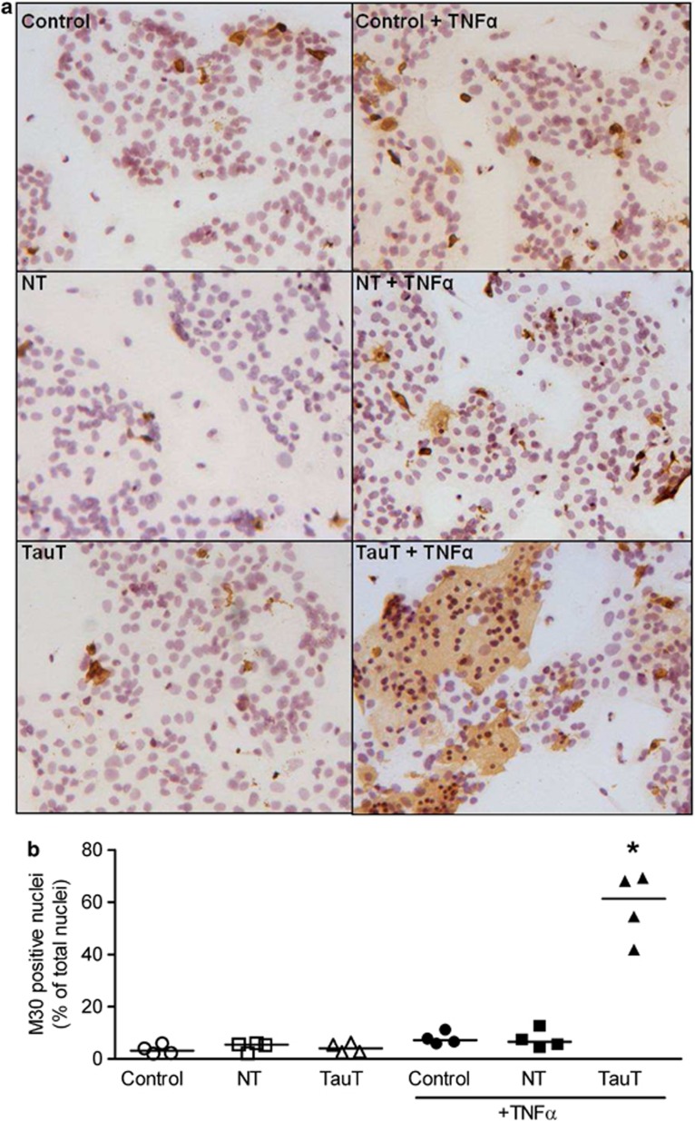
Figure 4.
TauT-deficient cytotrophoblast cells are more susceptible to TNFα-induced apoptosis. (a) Detection of apoptosis in primary cytotrophoblast cells using positive staining for caspase-cleaved cytokeratin 18 (M30 CytoDEATH). Counterstained with hematoxylin. M30 staining (brown) appears in the cytoplasm of apoptotic cells. (b) Scatter plot of M30-positive cells expressed as a percentage of total number of nuclei (n=4, line represents the median). *P<0.05 Kruskal–Wallis with Dunn's post test. NT=non-targeting siRNA, TauT=SLC6A6-specfic siRNA, +TNFα=overnight treatment with 100 ng TNFα

