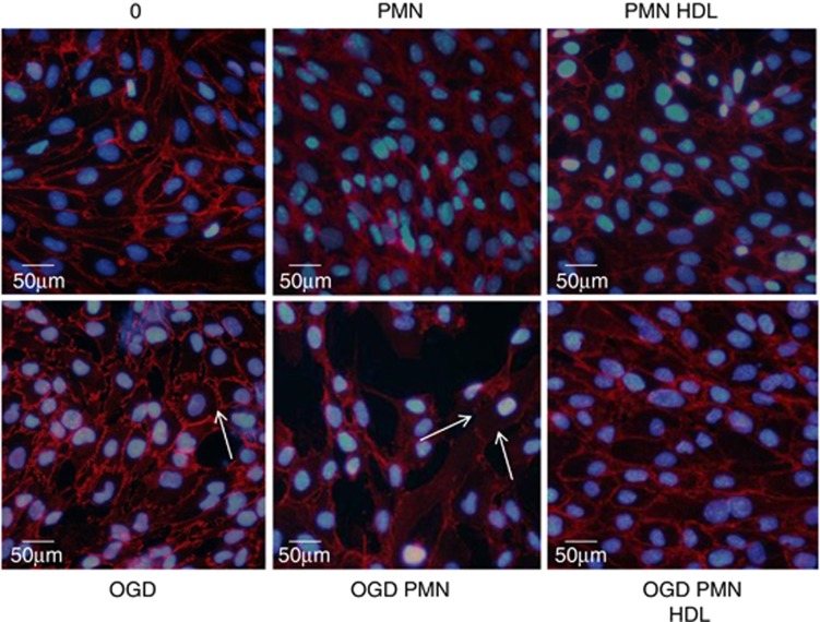
Figure 3.
Immunofluorescent staining of VE-Cadherin (red) after treatment with high-density lipoproteins (HDLs) (0.4 g/L)±polymorphonuclear neutrophils (PMNs) (1 × 10⁶ cells/mL) for 4 hours under normoxic or oxygen-glucose deprivation (OGD) conditions. Nuclei are stained with DAPI (blue). Results are representative of three independent experiments. The color reproduction of this figure is available at the Journal of Cerebral Blood Flow and Metabolism journal online.

