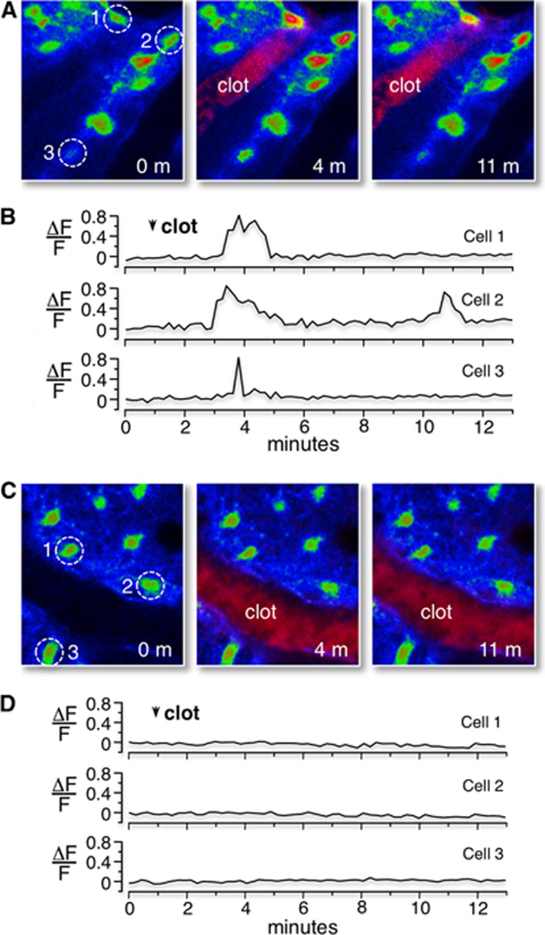
Figure 5.
Tail-vein injection of 2MeSADP induces Ca2+ increases in wild-type control astrocytes after photothrombosis, but not in astrocytes of IP3R2 KO mice. (A) Confocal images of the Ca2+ responses in wild-type control mice loaded with the Ca2+ indicator fluo4-AM before RB-induced photothrombosis, and 3 and 10 minutes after RB photothrombosis. Clotted vessel appears in the middle panel (red pixels with label). Ca2+ levels (fluo4 fluorescence) are color-coded (blue is lowest concentration, red is highest concentration). The Ca2+ levels of three cells (identified by dashed circles) are followed before and after 2MeSADP leakage across the blood–brain barrier. (B) Single-line traces of the Ca2+ responses from these three cells are plotted (ΔF/F). (C) Confocal images of Ca2+ response in IP3R2 KO mice before, and 3 and 10 minutes after RB photothrombosis. Imaging and presentation identical to wild-type mice. (D) Single-line traces of representative Ca2+ response of the cells identified in image panels.

