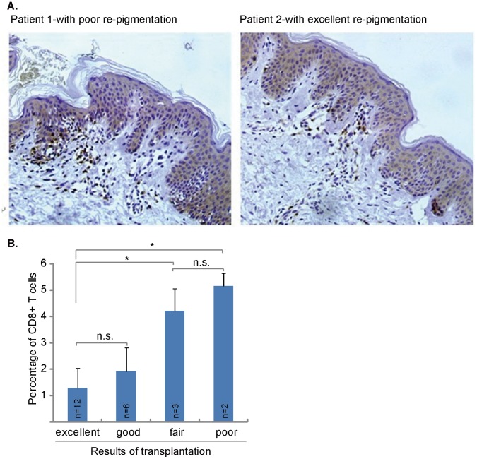
Figure 1. Perilesional CD8+ T cell infiltration is associated with the re-pigmentation efficiency of patients with autologous melanocytes transplantation.
Presence of CD8+ T cells in the perilesional skin of vitiligo lesions. Immunohistochemistry analysis of the perilesional skin of vitiligo patients revealed CD8+ T-cell infiltrations (DAB staining) (A). Patient 1 obtained 30% re-pigmentation after transplantation, patient 2 obtained 85% re-pigmentation after transplantation. Photos show the anti-CD8 mAb (brown) staining of two representative patients. Statistical analysis shows that the difference of CD8+ T cell infiltrating mainly exist between patients with fair/poor re-pigmentation and patients with excellent re-pigmentation (B). *p<0.05. n.s.: no statistical difference. Immunohistochemistry staining, original magnification ×200 (A and B).

