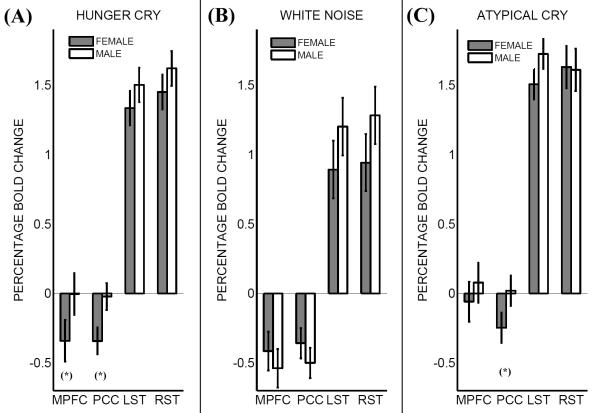
Figure 1.
Percentage BOLD (Blood Oxygenation Level Dependent) signal change versus rest in males and females in three conditions: (panel A) Hunger Cry, (panel B) White Noise, and (panel C) Atypical Cry. Asterisks denote significant differences between males and females’ percentage bold change (p<0.05). MPFC stands for dorsal medial prefrontal cortex, PCC for posterior cingulate cortex, LST for left superior temporal gyrus (Heschl’s gyrus), and RST for right superior temporal gyrus (Heschl’s gyrus). For precise coordinates of regions, see Table 1. Error bars are standard errors. Deactivation in dorsal medial prefrontal cortex and posterior cingulate cortex during hunger cry – distinctive of the mind-wandering interruption – took place only for the female group, but not for the male group. During atypical cry, this gender difference was significant only in posterior cingulate cortex, but not in dorsal medial prefrontal cortex.

