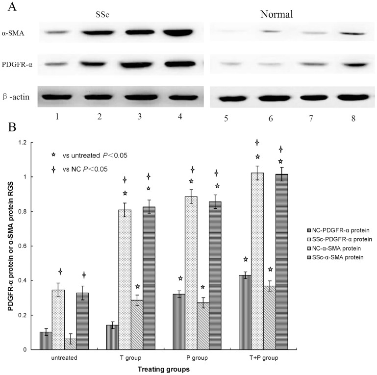
Figure 2. A: PDGFR-α and α-SMA protein expression stimulated with TGF-β1 and PDGF-AA.
SSc and normal skin fibroblast cultures were left untreated (columns 1, 5), costimulated with 10 ng/ml TGF-β1 plus 25 ng/ml PDGF-AA (columns 2, 6), stimulated with 10 ng/ml TGF-β1 (columns 3, 7) and stimulated with 25 ng/ml PDGF-AA (columns 4, 8). PDGFR-α protein and α-SMA protein expression was determined by WB. β-actin served to normalize expression of protein. B: Quantification of the data. RGS for PDGFR-α protein and α-SMA protein expression by cultured fibroblasts from SSc skin lesions and normal skin left untreated or stimulated with 10 ng/ml TGF-β1 (T group), 25 ng/ml PDGF-AA (P group), or 10 ng/ml TGF-β1 plus 25 ng/ml PDGF-AA (T+P group). PDGFR-α protein and α-SMA protein expression is up-regulated in SSc fibroblasts stimulated with TGF-β1 and PDGF-AA.

