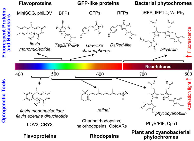
Fig. 1.
A diversity of the chromophores in the major groups of currently available fluorescent proteins, fluorescent biosensors, and optogenetic tools developed for biotechnological applications are shown. The upper part of the figure shows the chemical structures of flavin mononucleotide, TagBFP-like, GFP-like, DsRed-like and biliverdin chromophores for the respective fluorescent proteins and biosensors derived from flavoproteins (MiniSOG8, phiLOV9), GFP-like proteins (BFPs, GFPs, RFPs)2, 3, and bacterial phytochromes (iRFP10, IFP1.411, Wi-Phy12). The lower part of the figure shows the chemical structures of flavin mononucleotide, retinal and phycocyanobilin chromophores for the respective optogenetic tools derived from flavoproteins (LOV214, CRY215), rhodopsins (channelrhodopsins16, halorhodopsisns16, OptoXRs17), plant and cyanobacterial phytochromes (PhyB/PIF19, Cph118). The chromophores are shown in their protein-linked forms. A color scale presents the wavelength range of fluorescence emission for the fluorescent proteins and biosensors, and the wavelength range of the activation/de-activation light for the optogenetic tools.

